 Central Florida Health and Wellness Magazine Health and Wellness Articles of the Villages
Related Articles


State-of-the-Art Imaging
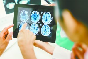 At ONC, patients with brain tumors undergo the latest imaging tests available. For tumors close to the speech or movement areas of the brain, functional MRI may be performed to identify their proximity to the tumor. Diffusion tensor imaging or “DTI” is another contemporary imaging technique that may be used for brain tumor surgery planning. This type of scan highlights the axons, or tracts, in the brain that connect one area to another. Together these imaging sequences aid the surgeon in creating a surgical gateway to be used during the operation thus maximizing safe tumor removal.
Intra-Operative Image Guidance: “Neuro-Navigation”
Stereotactic surgery, also called stereotaxy, is an advanced surgical technique used to treat be-nign and malignant tumors in the brain, skull base and spine utilizing computer-based, three- dimensional image guidance. “Neuro-navigation has revolutionized neurosurgery resulting in shorter operative times, smaller incisions, greater accuracy and less injury to the normal tissues around the tumor,” explained Dr. Jacob Freeman, newly acquired neurosurgeon at Ocala Neuro-surgical Center.
Awake Brain Tumor Surgery for Speech and Movement Preservation
Awake brain tumor surgery is reserved for patients with brain tumors involving eloquent areas of the brain
responsible for speech and movement. Using a strategy known as cortical and subcorti-cal mapping, the vital functional areas of the brain can be identified during surgery, along with their proximity to the tumor. Real time feedback from the awake patient can then be used during tumor removal to warn the surgeon if these areas are being affected by the surgery and prevent severe, permanent damage. Studies have shown that preservation of speech and movement re-sults in improved quality of life and better outcomes for patients. This is the goal for each of our patients at ONC.
“Using the latest technologies for neuro-navigation and brain mapping along with awake tech-niques, I’m able to offer safer, more complete tumors resections, which lead to better outcomes for my patients,” continued Dr. Freeman.
Dr. Freeman has specialized fellowship training in stereotactic brain and skull base tumor surgery, and is a published contributor to medical journals on various forms of this and other neuro-surgical procedures.
These state-of-the-art techniques are used to treat a variety of benign and cancerous lesions, in-cluding:
Metastatic brain tumors – Also called secondary brain tumors, these result from the spread of cancer cells from an area of the body to the brain and/or spine. Metastatic brain tumors are five times more common than primary brain tumors (tumors that originate in the brain).
Meningioma – Meningiomas are one of the most common types of primary brain tumors, ac-counting for nearly one-third of cases. These tumors appear among the meninges, the protective layers of tissue between the brain and skull. In most cases, they develop slowly, and may go un-detected for years before causing symptoms.
Glioma – Another common type of primary brain tumor, a glioma grows within the brain and can mix with or destroy healthy tissue, causing various symptoms, depending on the location and size. These tumors display a range of behaviors from slow-growing to fast and highly aggressive. In many cases, additional treatment including radiation and chemotherapy are administered after surgery.
Pituitary adenomas – Pituitary adenomas are benign, slow-growing tumors affecting the pituitary gland, a pea-sized organ located in the center of the brain behind the sinuses. Since the pituitary gland regulates hormones, a pituitary adenoma may cause serious symptoms and illness. Treat-ment involves the
expertise of a neurosurgeon and endocrinologist (hormone specialist).
Craniopharyngioma – A benign tumor that can encroach upon and adhere to the pituitary gland, optic nerve and other areas. Craniopharyngiomas most often affect children and adults over 50. They compose about two to four percent of primary brain tumors.
Chordoma – This slow growing, locally aggressive tumor most frequently forms at the skull base or within the bottom of the spine. Chordomas have a propensity to recur and are frequently treat-ed with surgery and a special type of proton radiation.
Hemangioblastoma – This is a rare variety of benign tumor that grows slowly within the menin-ges and blood vessels of the brain and spinal cord. A hemangioblastoma may not cause symp-toms until it ruptures and causes bleeding.
Tumor symptoms vary with type, location and size, but most often include headache, facial pain or numbness, dizziness, vision problems, ringing in the ears and hearing loss. Pituitary tumors can cause a host of symptoms and diseases associated with hormone imbalance. Using image guidance, Dr. Freeman performs more precise, safe and complete brain tumor resections in less time, leading to faster recovery for patients.
Ocala Neurosurgical Center
352-622-3360
www.OcalaNeurosurgicalCenter.com