By Mary Barber MD, The Skin Cancer Center of Central Florida –
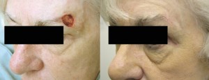
 This unique method of removing skin cancer and preserving normal surrounding skin has actually been available since the 1930’s. Dr Fredric Mohs of Wisconsin used a zinc chloride paste on the cancer and then removed the chemically fixed tissue and examined it under the microscope. This took 24 hours for each stage. Therefore, this method of skin cancer removal did not become practical until the late 70’s when the fresh frozen tissue technique was developed.
As the skin cancer epidemic exploded, more dermatologists became proficient in this technique and 2 methods of training became available. The first was established in 1967, the American Mohs College of Surgeons. A dermatologist could train under another Mohs surgeon for 1-2 years and become a Fellow of the Mohs College of Surgeons. Dermatology residencies grew to incorporate a Mohs surgeon to train the residents for their 3 years. Many dermatologists had become very proficient in the Mohs procedure in their residencies and therefore in the early 90’s , the American Society of Mohs Surgeons (ASMS) was founded. To become a Fellow, 75 surgical cases had to be presented and a certifying exam passed. In 1995, I became a Fellow of the ASMS. I have performed over 15,000 cases of Mohs surgery.
THE MOHS MICROGRAPHIC PROCEDURE
1) In the dermatology office, the tumor is outlined with a marker by the surgeon and the area is anesthetized with a local anesthetic.
2) The cancer is excised from the normal appearing skin and a temporary bandage is placed. The patient now waits for the tissue to be processed.
3) The specimen is taken to the lab in the office and is inked and mapped ( “a graph” is made)
4) It is now flattened and frozen at 26 degrees Celsius. This is the difference between Mohs and a typical frozen section! Mohs histotechnicians make sure that 100% of the margins are able to be visualized by the pathologist. (who is always the Mohs surgeon)
5) The specimen is sliced very thinly and stained.
6) In about 40 minutes the slides are ready. The Mohs surgeon reads the slides under the microscope. (This is the “micro” part)
7) If the tumor is all out, then the defect can be closed.
8) If there is still tumor – then the exact location can be pinpointed and another specimen is taken and the process is repeated (Stage 2) 80% of the time, the tumor is removed in 1 or 2 Stages. This is the national standard. Many Mohs surgeons are trained to do plastic surgery closures of the defects. I have done thousands of skin grafts and many other cosmetic closures.
The advantages of Mohs surgery are:
1) Only cancerous tissue is removed, extra margins of normal skin are not excised like they are in routine excisions of skin cancer.
2) The patient knows that day whether the skin cancer is all out.
3) The resultant surgical scar is smaller.
4) The cure rate is 99% for tumors that have never been operated on before and 95% if this was a recurrent tumor.
5) It is much less expensive than radiation or excision with standard frozen sections.
So why isn’t Mohs micrographic surgery used for all skin cancers?
There are 2 other well proven surgical treatments that give excellent cure rates and are less expensive – Curettage and Electrodessication X 3 and Excisional surgery. In the next article, I will review these and other methods of skin cancer treatment.
Patients routinely ask me to perform Mohs surgery on their skin cancers. I am happy to oblige if the tumor meets the qualifications adopted by Medicare and the American Academy of Dermatology. As this treatment is time intensive and maybe more expensive than the 2 other surgical techniques, guidelines have been set up so as not to overuse Mohs surgery.
The Main Indications of Mohs Micrographic Surgery are:
1) Recurrent skin cancers
2) Patients who are immunosuppressed (ie Kidney transplant patients)
3) Patients younger than 40 years of age
4) Skin cancers with aggressive pathology reports (basically they have roots than invade or are a type that are known to be able to spread throughout the body)
5) Cancers greater than 2 cm in diameter.
6) Cancers found on the eyelids, ears, central face, nose, hands, feet, and genital area ( Yes, skin cancer can occur there! Getting a total body skin exam is a really good thing!)
Approximately 25% of Floridians will get a skin cancer. Most will have more than one. It is important to know your treatment options and exercise them!
At The Skin Cancer of Central Florida, each patient is unique and their treatment plan should be also. Our motto is Experience Makes the Difference. Visit us and see!
About Dr Barber
Dr. Mary F. Barber has performed over 15,000 Mohs procedures. She limits her practice to the treatment of proven skin cancer patients. New patients who need skin checks are welcomed at the Skin Cancer Center and should make an appointment to see Dr. Corwin, Nurse Practitioner Mary Jane Oates, or Physician Assistant Theresa Saleh. No referral is needed.
Skin Cancer Center of Central Florida | 352-259-6553 | www.skincancersurgery.net
Check Also
Recurrent UTIs: Addressing the Risk of Antibiotic Resistance
Urinary tract infections (UTIs) are common bacterial infections that affect millions of individuals worldwide each …
 Central Florida Health and Wellness Magazine Health and Wellness Articles of the Villages


