 Central Florida Health and Wellness Magazine Health and Wellness Articles of the Villages
Related Articles


 If your doctor has said you need a neuroimaging exam, you certainly have questions about what it is and what to expect.
Herein we will cover the general principles of neuroradiology.
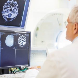
Neuroradiology is a subspecialty within the practice of radiology that addresses conditions affecting the brain, skull, spine, spinal cord, neck and central nervous system. Neuroradiologists are doctors who diagnose a huge range of disorders, including congenital malformations, genetic conditions, trauma, stroke, tumor, aneurysm, neurodegenerative diseases, MS, infection, metabolic disorders and others. The technologists are the highly trained people who perform the imaging exams, while the neuroradiologists are the physicians who interpret the results, form a diagnosis and work with your referring clinician to help stage, and often perform, any treatment.
“Subspecializing in neuroradiology requires a minimum of 11 years of post-graduate education and training,” says neuroradiology subspecialist, Dr. Edilberto Alvarez. “It is one of the most complex and fascinating radiological subspecialities, one in which advanced image guidance plays a crucial role in both diagnosis and treatment.”
While having a neuroimaging exam may seem a little scary at first, these tests are painless, noninvasive and capable of revealing even tiny abnormalities, many of which can be treated using minimally-invasive procedures that can safely restore health and function. RAO’s team of neuroradiologists interpret images created by leading-edge technologies, including advanced x-ray and CT to provide an accurate diagnosis and assist referring clinicians by recommending the best testing modalities for each patient’s symptoms and history. Neuroradiologists also treat many of these conditions, such as arteriovenous malformations, stroke, spinal compression fractures and others, without the risk, complications or long healing times associated with surgery.
Magnetic resonance imaging, or MRI, and computed tomography, or CT, are two of the most important technologies used in neuroradiology, as they create highly detailed images of the brain, spine, blood vessels and related systems. MRI uses radio waves and a powerful magnet to create images without ionizing radiation. The exam involves lying down on an exam table, which slides into the imaging machine. You are asked to remain still for 30-45 minutes while a series of 3-D quality images is captured.
With a CT scan, you also lie down on an exam table, but this test takes only about 10 minutes. You may be asked to hold your breath for short periods while images are taken. X-ray imaging is another imaging tool used for procedures like catheter angiography to investigate blood vessels for aneurysm, plaque and other problems. Apart from an injection of contrast dye to highlight details, all of these tests are completely painless.
“Using image guidance, we treat medical problems using tools like catheters, stents and injections,” says neuroradiologist Dr. Ralf R. Barckhausen. “Many conditions that used to require open surgery, such as an aneurysm, subarachnoid hemorrhage and stroke, can be treated with minimal invasiveness. We can also use image guidance to perform pain management procedures like epidural injections and nerve root blocks to help provide relief from many chronic pain syndromes.”
By getting an accurate diagnosis, your care team can work together to develop the right treatment plan to help get you back to health, vigor and better quality of life.
Radiology Associate of Ocala
RAOcala.com
352-671-4300