 Central Florida Health and Wellness Magazine Health and Wellness Articles of the Villages
Related Articles


By Amanda M. Burns, M.D.
Munroe General Surgery, General and Breast Surgery
 The diagnosis of breast cancer is a life-changing event for many women. Those four words – “You have breast cancer” are met with a million emotions, from not only the woman facing the diagnosis, but also her loved ones. Unfortunately, this cancer is far too common, affecting over 250,000 women every year. In addition to patients with a cancer diagnosis, thousands of women are diagnosed with precancerous lesions that not only require similar surgical workups, but also lead to an increased risk of developing cancer in the future. But the great news is that for many women, a chance of cancer cure is attainable! The majority of women diagnosed with breast cancer will be cured of their cancer after treatment, with early stage cancers having a nearly 100% chance of cancer cure! Thanks in part to new and improved chemotherapy agents and advancement in radiation treatments, women are no longer faced with an incurable disease.
In addition to improvements in therapy, early detection is a key to these incredible outcomes! For this reason annual screenings are paramount for any women over the age of 40. That is why we focus on educating women on the importance of breast self-exams and knowing your body. But what are the important things to look for, what warning signs should women be aware of, and when should a women seek further evaluation by her physicians? What features on imaging are clinicians looking for, and what findings warrant further evaluation? My hope is that at the end of this article, I will have answered many of these questions and given the women of this community the power to take charge of their health and an understanding of breast disease.
Let’s start with breast examination. Although not absolutely required, women are encouraged to preform monthly breast self-exams. For the most part, this is to establish an understanding of your body and to know what is normal for you.
Everyone’s body is different, and everyone’s breasts are different. That is why understanding what is normal for you is important. It is when something changes that you should be aware. A monthly exam, performed at the same time of your menstrual cycle, is important to appreciate these changes. Additionally, every woman should have a yearly exam performed by her clinician. Changes to be aware of include a new lump that has not been present before, particularly if it is hard, painful, growing, or associated with overlying skin changes. Nipple changes can also be a sign of an underlying process. These include nipple retraction that is new; nipple discharge, particularly if it is only in one breast or bloody; or skin changes of the nipple. These are the same things your primary physician looks for when they perform a clinical breast exam.
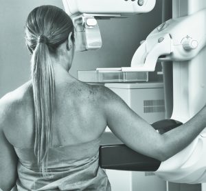
Imaging is often the next step in evaluation of any patient with concerns. It is also our best tool for screening, as many cancers and precancerous lesions are not palpable and only found on imaging. There are two mainstays of breast imaging – ultrasound and mammogram. For women over the age of 40, a yearly mammogram is recommended. Mammograms are an excellent tool for looking at various changes within the breast. They work by compressing the breast and using x-rays to take an image of the underlying breast tissue. The amount of radiation delivered with each mammogram is very limited, and any risk from the radiation exposure is very small. Mammograms allow clinicians to identify masses, as well as more subtle changes, such as micro-calcifications or architectural distortions. While no imaging modality can accurately diagnose cancer without a tissue biopsy, these findings on a mammogram increase the suspicion of an underlying process – whether precancerous or cancerous. Women with findings such as these on mammogram will often undergo further testing with either additional imaging or with biopsy to make a full diagnosis.
Often times, women with concerning findings on a routine mammogram will be asked to come back to have another mammogram performed. Many women ask why it is necessary to undergo the same imaging again. The answer is simple – for women without any concerns, a screening mammogram is performed. This type of mammogram utilizes two standard views of each breast. These two routine views are great for evaluating the full breast, and are consistent across the country. Often, these can be performed without a physician’s authorization, so for women without a primary care physician, a screening mammogram can be obtained without an order from a clinician.
For women with a palpable mass, or a concerning finding on screening mammogram, further imaging may be needed to fully evaluate the area of concern. This is when a diagnostic mammogram is performed. The views, or techniques, vary depending on where the area is, and what it looks like on the original screening mammogram. Various techniques may be applied, such as increased compression of the breast in the area of concern, as well as changing the angle at which the mammogram is taken. Many radiology centers will perform a biopsy at the time of a diagnostic mammogram if warranted, thus eliminating an extra visit to the radiology center, and expediting the diagnosis.
While mammograms are a great tool for evaluating breast disease, they have their limitations. For women with dense breasts, often women under the age of 40, mammogram is not always the best option. For these women, ultrasound may be performed alone, or in conjunction with a mammogram, to allow for further evaluation of the breast tissue. Ultrasound is a great tool for evaluating cysts seen on mammogram, and determining if these cysts are benign appearing or warrant further imaging or biopsy. Additionally, palpable masses are further evaluated with ultrasound to differentiate between cystic and solid masses. Just as with mammograms, certain features are more indicative of an underlying process that requires further testing.
For any woman with concerns regarding her breast, whether it is a new lump, pain, discharge, or another concerning finding, reaching out to your physician care team is always the best option. It is never a bad idea to have your concerns evaluated by your trusted clinician. My goal is to provide the wonderful women of this community an increased knowledge of breast disease. With this increased understanding, women can take charge of their health, and can be empowered to seek further answers. Ultimately we can find and treat any breast cancer while it is in an early stage, and give every woman with the diagnosis of breast cancer a chance at a 100% cure.
Dr. Amanda Burns is a General and Breast Surgeon with Munroe Medical Group and has offices located in Ocala and Lady Lake. She can be reached at 352-671-1202, and is currently accepting patients and can offer next day appointments. She can also be reached by email at amanda_burns@munroregional.com. If you have any questions, comments, or concerns please do not hesitate to call or email.
Perform a monthly self-examination
Women should perform a self breast-exam each month and any changes or abnormalities should be discussed with a doctor or physician.
1) In the Shower
Using the pads of your fingers, move around your entire breast in a circular pattern moving from the outside to the center, checking the entire breast and armpit area. Check both breasts each month feeling for any lump, thickening, or hardened knot. Notice any changes and get lumps evaluated by your healthcare provider.
2) In Front of a Mirror
Visually inspect your breasts with your arms at your sides. Next, raise your arms high overhead. Look for any changes in the contour, any swelling, or dimpling of the skin, or changes in the nipples. Next, rest your palms on your hips and press firmly to flex your chest muscles. Left and right breasts will not exactly match—few women’s breasts do, so look for any dimpling, puckering, or changes, particularly on one side.
3) Lying Down
When lying down, the breast tissue spreads out evenly along the chest wall. Place a pillow under your right shoulder and your right arm behind your head.
Using your left hand, move the pads of your fingers around your right breast gently in small circular motions covering the entire breast area and armpit. Use light, medium, and firm pressure. Squeeze the nipple; check for discharge and lumps. Repeat these steps for your left breast.
Source: Nationalbreastcancer.org
Cancer.org
MRMC
1908 SE 18th Avenue, Bldg 200, Ocala, FL 34471 (352) 671-1201